Contact
|
Research
|
Publications
|
Lab Members
| Gallery |
Funding
|
Protocols
|
Join the Lab!
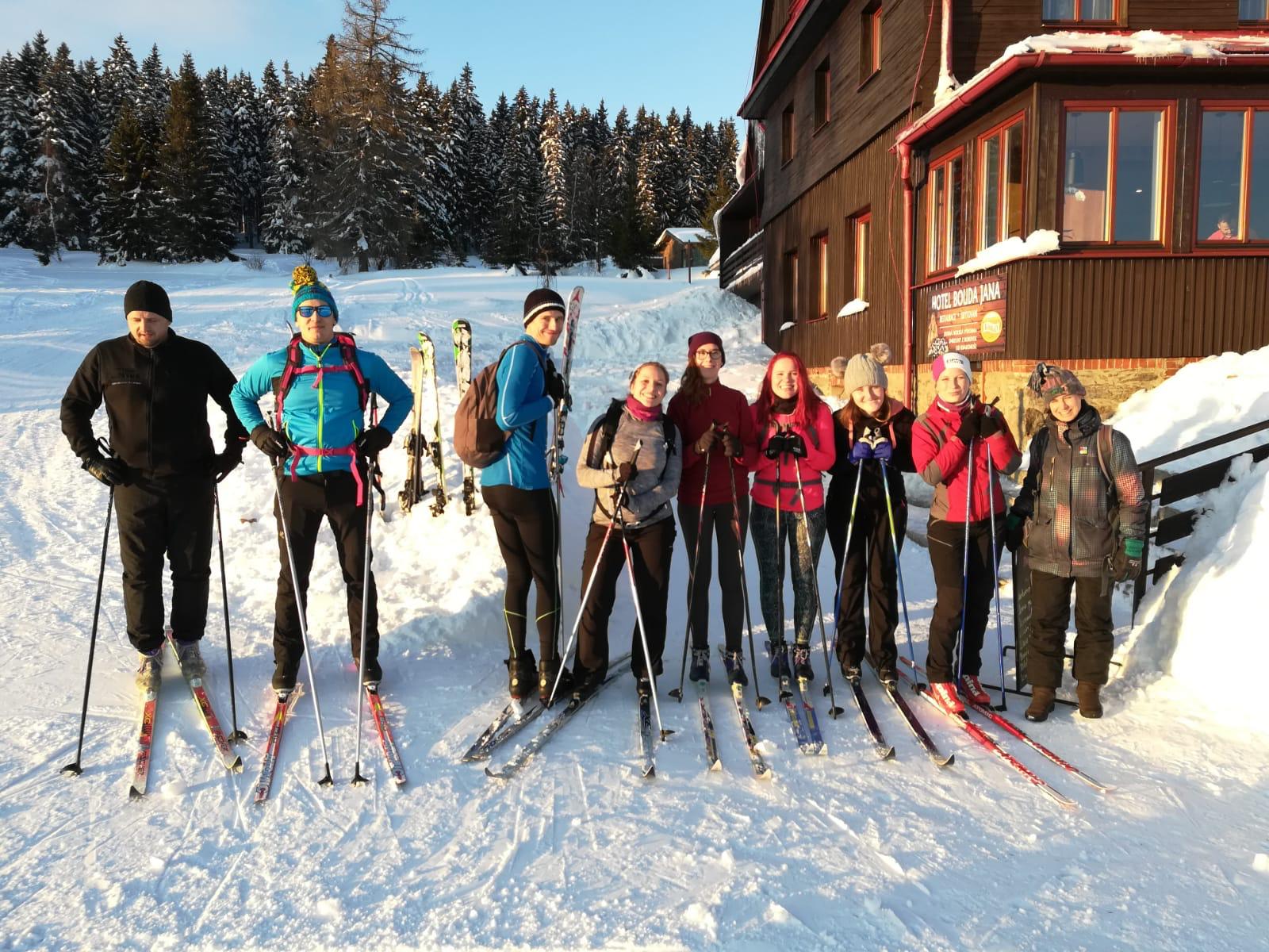
Skiing trip in Krkonose
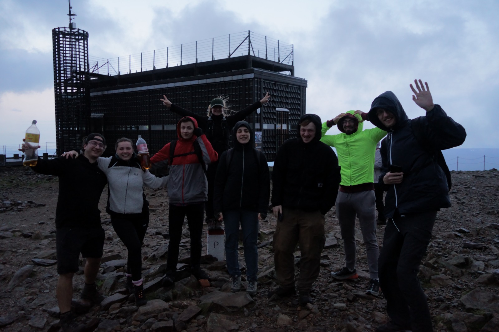
LAB retreat
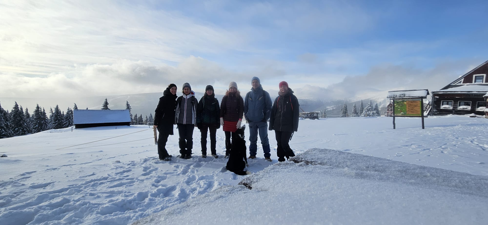
LAB retreat II
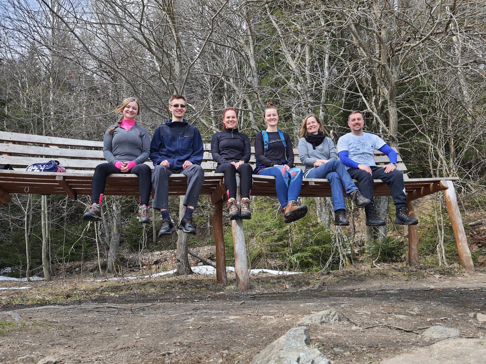
LAB retreat III - Czech Railways