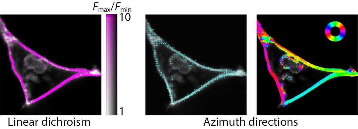
Fluorescence-detected linear dichroism microscopy is a useful technique for observing various molecular processes in living cells. It also allows determining the orientation of fluorescent molecules with respect to cellular structures. However, until recently, interpreting images acquired by linear dichroism microscopy has been rather laborious, time-consuming, and unreliable.
A research team led by Josef Lazar from IOCB Prague and the Institute of Microbiology of the CAS has developed a set of novel open-source ImageJ-based software tools to make visualization and data interpretation more accessible. The article describing their work has now been published in Communications Biology.
The new software tools for processing data acquired by linear dichroism microscopy are applicable to images of a wide range of samples, including those of model synthetic lipid vesicles, but also of living cells with complex shapes, labeled genetically by fluorescein proteins. The study contributes to making polarization microscopy more accessible or even a mainstream tool for biological imaging in the near future.
Read the paper:
- Bondar, A.; Rybakova, O.; Melcr, J.; Dohnálek, J.; Khoroshyy, P.; Ticháček, O.; Timr, Š.; Miclea, P.; Sakhi, A.; Marková, V.; Lazar, J. Quantitative linear dichroism imaging of molecular processes in living cells made simple by open software tools. Commun. Biol. 2021, 4, 189. https://doi.org/10.1038/s42003-021-01694-1